Broadly speaking, brain is the anatomical structure and mind is the physiological (functional) component.
In another simple manner, brain can be described as the hardware like a computer hardware and mind can be described as the software of a computer.
When we talk about Neuroanatomy then it means study of the brain, spinal cord, cranial nerves , spinal nerves , nerve plexuses, dermatomes, muscles and joints supplied by these nerves. At the outset it seems very tough to master neuroanatomy, but it is very interesting and easy to learn.
Functionally , the nervous system is divided into the somatic nervous system, which controls the voluntary activites and the visceral ( autonomic) nervous system, which controls involuntary activities.
Anatomically, the nervous system can be divided into 2 subdivisions:
CNS (Central Nervous System) & PNS (Peripheral Nervous System).

CNS consists of brain & spinal cord.
PNS consists of cranial nerves & spinal nerves. The peripheral nervous system consists of 12 pairs of cranial nerves and 31 pairs of spinal nerves , and their associated ganglia.

As the basic building materials of a house are brick and cement, similarly the Nervous system is made by the Neurons and Neuroglia.
Neuron is the basic unit of nervous system.
Neuroglia are non-neuronal cells and these are of three types: Astrocytes, Oligodendrocytes and Microglia.
Neuron or nerve cell is the basic functional unit of the nervous system. There are about 100 billion neurons in central nerovous system. They are supported by glial cells. They are present in brain and spinal cord and glial cells outnumber neurons (10:1).
Neurons are the structural and functional units of the nervous system.
Neuronons consist of cell bodies ( perikaryon or soma) with dendrites and axon.
Dendrites ( dendron means tree) are short and highly branched and carry impulses toward the cell body.
Axons are usually single and long, have fewer branches ( collaterals), and carry impulses away from the cell body.
Myelin is the fat like substance forming a sheath around the axons of certain nerve fibers. It is formed by Schwann cells in the PNS and oligodendrocytes in the CNS.
Neuroglial cells or Glial cells are of three types: astrocytes, oligodendrocytes and microglia. Astrocytes and Oligodendrocytes can be classified together as Macroglia. Macroglia is dervived from ectoderm. Astrocytes regulate the ionic environment & reuptake of neurotransmitters. They form the blood brain barrier. There are two broad classes of astrocytes: protoplasmic and fibrous. Astrocytes provide structural support to nervous tissue and act during development as guidewires that direct neuronal migration. They also maintain appropriate concentration of ions such as Potassium ions within the extracelluar space of brain & spinal cord.
Astrocytes form a covering on the entire CNS surface & proliferate to aid in repairing to aid in repairing damaged neural tissue. These reactive astrocytes are larger, are more easily stained, and can be identified in histological sections because they contain a characteristic, astrocyte-specific protein: glial fibrillary acidic protein (GFAP). Chronic astrocytic proliferation leads to gliosis, sometimes called glial scarring.
Oligodendrocytes form the myelin sheath around neurons in central nervous system. ( In peripheral nervous system , i.e., in case of spinal nerves, the Myelin sheath is formed by the Schwannn cells).
Oligodendrocytes predominate in white matter; they extend arm-like processes which wrap tightly around axons.
Microglia or microglial cells are the macrophages or scavengers of the CNS & do the immune surveillance of the CNS.
Nerve cells convey signals to one another at synapses. Chemical transmitters ( or Neurotransmitters) are associated with the function of the synapse: excitation or inhibition. A neuron may receive thousands of synapses, which bring in information from many sources.
Brain anatomy can be described under following headings: cerebrum, cerebellum and brainstem. Cerebrum consists of two cerebral hemispheres which are connected to each other with commissural fibers. The largest bundle of commissural fibers is corpus callosum, which connects the two cerebral hemispheres.
Cerebellum is situated on posterior aspect of brain, below the cerebrum and behind brain stem
Brain stem comprises Midbrain, Pons and Medulla oblongata.
Brain constitutes about 2% of the body weight but receives about 18-20% of cardiac output. It controls all the functions of the body. It is of great interest to everyone. The human brain is very much different from the brain of other creatures in view of its immense capacity. A large surface area of the neural tissue is contained inside the cranium or skull. It is possible because of large number of infoldings which take the shape of sulci and gyri. The part which caves in is called the sulcus and the elevated part is called gyrus.
The neuroanatomy is very interesting and lot of advancements have occurred in the understanding of the microneurosurgical anatomy of the brain.
Almost all body parts of the body are represented in the cerebral hemispheres. Different areas of the cerebral hemisheres have been assigned different functions.
Three membranes cover the brain and spinal cord, these are known as meninges. The innermost is Piamater, middle layer is Arachnoid layer and the outermost is the thickest layer, the Duramater. The Dura is also called the pachymeninx, and the arachnoid and pia are called the leptomeninges. the dura mater is tough, fibrous sheath and is continuous with the spinal dura.
The arachnoid is a thin, transparent sheath separated from the underlying pia by the subarachnoid space, which contains cerebrospinal fluid ( CSF).
Brain floats inside a fluid called cerebrospinal fluid (CSF). CSF is contained between Arachnoid layer and Piamater.
Cerebral hemispheres, corpus callosum, brain stem , cerebellum are contained inside a hard bony structure known as cranium or skull. To protect the soft brain against the hard bony structures, there are wide CSF spaces at the base of the brain, known as CSF Cisterns.
Spinal cord is contained in the vertebral column or spine. Spinal cord is about 45 centimeter in length and about 30 grams in weight. Spinal cord is continuous with the medulla at its upper end. Conus medullaris is the lower or inferior end of the spinal cord.In adults, the conus ends at the lower border of the L1 vertebra.
Brain and spinal cord together is known as central nervous system. The cranial nerves and spinal nerves constitute the peripheral nervous system.

Weight of brain in an adult is about 1,300 - 1,400 Grams (Approximately 1.4 kg).
Weight of brain in a newborn is about 350 - 400 Grams.
Brain constitutes 2% of total body weight.
Intracranial contents by volume (1,700 ml, 100%):
brain = 1,400 ml (80%);
blood = 150 ml (10%);
cerebrospinal fluid = 150 ml (10%)
Average number of neurons in the brain = 100 billion
Average number of glial cells in brain = 10-50 times the number of neurons
Rate of production of CSF = 0.35 ml/min (500 ml/day)
% blood flow from heart to brain = 15-20%
Blood flow through whole brain (adult) = 750 ml/min
% brain utilization of total resting oxygen = 20%
Cerebral blood flow = 55 ml/100 g brain tissue/min
Cerebral cortex on surface can be divided into four major lobes- Frontal, Parietal , Temporal and Occipital Lobes.
To summarise, the weight of brain in an adult is approximately 1.4 kg and it appears as a soft structure. It is covered inside three coverings : the outermost layer is called duramater, the middle layer is called Arachnoid layer and the innermost layer is Pia mater. In between Arachnoid and Pia mater there is a space called Subarachnoid space which contains Cerebrospinal fluid ( CSF ).
 (Fig Source: www.shortfacts.com)
The cadaveric brain without blood vessels looks like this picture. It has various infoldings which increase the surface area of the brain. There are various sulci and gyri.
 (Fig source en.wikipedia.org)
 (Fig Source Morphonix)

To understand neuroanatomy, one should begin with practice of drawing lateral surface of the brain.
 This figure simplifies the various parts inside the brain . The cerebrum and cerebellum are situated on the stem , known as brain stem. Brain Stem consists of Mid Brain, Pons and Medulla Oblongata.


 Actual look of a sagittal section of brain of a human cadaver showing Mid sagittal structures of the brain. An understading of this picture will help you to read a sagittal image of the MI of the brain. It shows corpus callosum, cerebellum, Midbrain, Pons and Medulla Oblongata.Corpus callosum connects two cerebral hemisheres.
 (Fig source: www.webmd.com)
This picture gives an idea about the skull ( Cranium), brain ( Cerbral hemispheres and Cerebellum), and spinal cord.

It is the mid sagittal section image : Below the corpus callosum is lateral ventricle and below the Fornix is third ventricle . The Floor of the third ventricle shows pituitary stalk. This picture gives an idea about the relation of the brain to the orbit, location of pituitary gland to the nasal cavity, location of the brain stem.
If someone has an idea about the axial , coronal and sagittal sections of the brain it becomes easy to interpret the CT scan and MRI of the brain.
Spinal cord is the continuation of the lower part of brain stem, i.e., medulla oblongata. Medulla oblongata ends at foramen magnum, i.e., an opening at the posterior end of skull.
Almost all the functions are regulated by the brain and some examples are motor function, speech, vision,etc. Different areas of the brain regulate different functions.
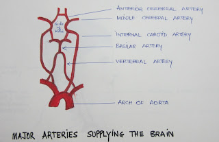
Arterial supply of the brain can be described as anterior circulation and posterior circulation. the anterior circulation is contributed by internal carotid artery and posterior circulation is contributed by vertebral arteries. Internal carotid artery is branch of common carotid artery and vertebral artery is a branch of subclavian artery.
The vertebral arterial system supplies the brain stem, cerebellum, parts of thalamus.
The internal carotid artery supplies blood to remainder of the brain (about 85%).
Circle of Willis is a circle of arteries at the base of the brain that gives rise to all the major blood supply to the brain. The circle of Willis is named after the English neuroanatomist Sir Thomas Willis. It is formed by two anterior cerebral arteries which are in communication with each other anteriorly through one anterior communicating artery. There are two posterior communicating arteries which are connected to the posterior cerebral arteries, the terminal branches of basilar artery.


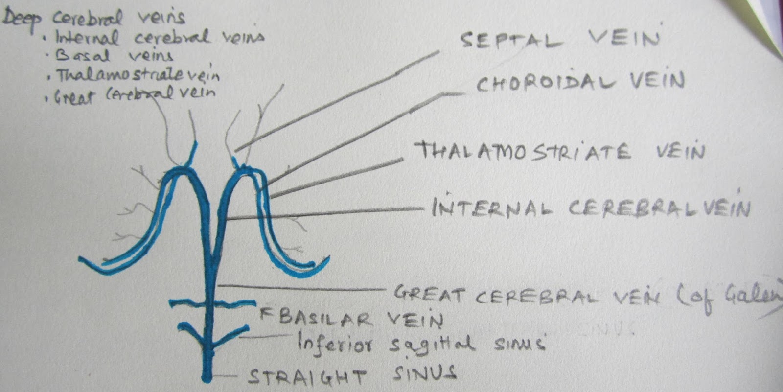
Venous system of brain
All the veins of the brain drain into Internal Jugular Vein which drains into right atrium of the heart. But the venous drainage of brain is different from other structures of the body. The veins of the brain can be described as superficial and deep venous systems of the brain.
Superficial venous system of the brain

Deep Venous System of the Brain

Cranial nerves
There are 12 pairs of cranial nerves
Cranial nerve I Olfactory nerve
Cranial nerve II Optic nerve
Cranial nerve III Oculomotor nerve
CN IV Trochlear nerve
CN V Trigeminal nerve
CN VI Abducens nerve
CN VII Facial nerve
CNVIII Vestibulocochlear nerve
CNI X Glossopharyngeal nrve
CN X Vagus nerve
CN XI Spinal Acessory nerve
CN XII Hypoglossal nerve
Cranial nerves 128 are sensory nerves, 5, 7 , 9 and 10 are mixed nerves and rest are motor cranial nerves.
( So, to know the type of cranial nerves , remember just two numbers 128, and 5790).
Contrary to the popular belief about difficulty in neuroscience and brain anatomy, it is very easy to understand the neuroanatomy. If someone aims to master the neuroanatomy, the way is very simple. Begin with understanding the basic anatomy of skull , brain , vertebral column and spinal cord in a step wise manner. I have drawn the pictures of the skull , brain and spinal cord to highlight the value of these basic facts.
This manner of learning is good for the medical students and neurosurgeons as well. The ability to draw the image of a skull helps in understanding the parts of skull . This helps in understanding neuroradiology and in the surgical planning also.

So, the first step is to see the skull. It is easy to identify frontal bone, parietal bone , temporal bone, occipital bone, orbit, and maxilla. locate nasal aperture in the frontal view. and pterion and zygomatic process in the lateral view of skull.


The first picture provides an idea about the three fossae of the inside of the cranium or skull. The anterior cranial fossa, middle cranial fossa and posterior cranial fossa. It is extremely important to know these three terms, as these three terms are frequently used by the experts in neuroanatomy and neurosurgery. This picture is a diagrammatic representation of the view of the skull base as seen from above after removing the skull cap. One should learn to draw this simple yet a very important diagram. Locate the greater wing of the sphenoid bone, anterior clinoid procees , petrous bone, posterior clinoid process and foramen magnum in this diagram. Rest of the three diagrams are are enlarged view of the base of the three intracranial fossae.
Just to make you aware about the inside of the cranium another simple diagram is drawn below . Identify and note the location of tent which divides inside the cranium into supratentorial and infratentorial compartments.

 This is the schematic transverse section of the spinal cord. Spinal cord is also bounded by three meningeal layers as that of brain. Subarachnoid space contains CSF. Ligamentum denticulatum is the extension of the Piamater in spinal cord.

In another simple manner, brain can be described as the hardware like a computer hardware and mind can be described as the software of a computer.
When we talk about Neuroanatomy then it means study of the brain, spinal cord, cranial nerves , spinal nerves , nerve plexuses, dermatomes, muscles and joints supplied by these nerves. At the outset it seems very tough to master neuroanatomy, but it is very interesting and easy to learn.
Functionally , the nervous system is divided into the somatic nervous system, which controls the voluntary activites and the visceral ( autonomic) nervous system, which controls involuntary activities.
Anatomically, the nervous system can be divided into 2 subdivisions:
CNS (Central Nervous System) & PNS (Peripheral Nervous System).

CNS consists of brain & spinal cord.
PNS consists of cranial nerves & spinal nerves. The peripheral nervous system consists of 12 pairs of cranial nerves and 31 pairs of spinal nerves , and their associated ganglia.

As the basic building materials of a house are brick and cement, similarly the Nervous system is made by the Neurons and Neuroglia.
Neuron is the basic unit of nervous system.
Neuroglia are non-neuronal cells and these are of three types: Astrocytes, Oligodendrocytes and Microglia.
Neuron or nerve cell is the basic functional unit of the nervous system. There are about 100 billion neurons in central nerovous system. They are supported by glial cells. They are present in brain and spinal cord and glial cells outnumber neurons (10:1).
Neurons are the structural and functional units of the nervous system.
Neuronons consist of cell bodies ( perikaryon or soma) with dendrites and axon.
Dendrites ( dendron means tree) are short and highly branched and carry impulses toward the cell body.
Axons are usually single and long, have fewer branches ( collaterals), and carry impulses away from the cell body.
Myelin is the fat like substance forming a sheath around the axons of certain nerve fibers. It is formed by Schwann cells in the PNS and oligodendrocytes in the CNS.
Neuroglial cells or Glial cells are of three types: astrocytes, oligodendrocytes and microglia. Astrocytes and Oligodendrocytes can be classified together as Macroglia. Macroglia is dervived from ectoderm. Astrocytes regulate the ionic environment & reuptake of neurotransmitters. They form the blood brain barrier. There are two broad classes of astrocytes: protoplasmic and fibrous. Astrocytes provide structural support to nervous tissue and act during development as guidewires that direct neuronal migration. They also maintain appropriate concentration of ions such as Potassium ions within the extracelluar space of brain & spinal cord.
Astrocytes form a covering on the entire CNS surface & proliferate to aid in repairing to aid in repairing damaged neural tissue. These reactive astrocytes are larger, are more easily stained, and can be identified in histological sections because they contain a characteristic, astrocyte-specific protein: glial fibrillary acidic protein (GFAP). Chronic astrocytic proliferation leads to gliosis, sometimes called glial scarring.
Oligodendrocytes form the myelin sheath around neurons in central nervous system. ( In peripheral nervous system , i.e., in case of spinal nerves, the Myelin sheath is formed by the Schwannn cells).
Oligodendrocytes predominate in white matter; they extend arm-like processes which wrap tightly around axons.
Microglia or microglial cells are the macrophages or scavengers of the CNS & do the immune surveillance of the CNS.
Nerve cells convey signals to one another at synapses. Chemical transmitters ( or Neurotransmitters) are associated with the function of the synapse: excitation or inhibition. A neuron may receive thousands of synapses, which bring in information from many sources.
Brain anatomy can be described under following headings: cerebrum, cerebellum and brainstem. Cerebrum consists of two cerebral hemispheres which are connected to each other with commissural fibers. The largest bundle of commissural fibers is corpus callosum, which connects the two cerebral hemispheres.
Cerebellum is situated on posterior aspect of brain, below the cerebrum and behind brain stem
Brain stem comprises Midbrain, Pons and Medulla oblongata.
Brain constitutes about 2% of the body weight but receives about 18-20% of cardiac output. It controls all the functions of the body. It is of great interest to everyone. The human brain is very much different from the brain of other creatures in view of its immense capacity. A large surface area of the neural tissue is contained inside the cranium or skull. It is possible because of large number of infoldings which take the shape of sulci and gyri. The part which caves in is called the sulcus and the elevated part is called gyrus.
The neuroanatomy is very interesting and lot of advancements have occurred in the understanding of the microneurosurgical anatomy of the brain.
Almost all body parts of the body are represented in the cerebral hemispheres. Different areas of the cerebral hemisheres have been assigned different functions.
Three membranes cover the brain and spinal cord, these are known as meninges. The innermost is Piamater, middle layer is Arachnoid layer and the outermost is the thickest layer, the Duramater. The Dura is also called the pachymeninx, and the arachnoid and pia are called the leptomeninges. the dura mater is tough, fibrous sheath and is continuous with the spinal dura.
The arachnoid is a thin, transparent sheath separated from the underlying pia by the subarachnoid space, which contains cerebrospinal fluid ( CSF).
Brain floats inside a fluid called cerebrospinal fluid (CSF). CSF is contained between Arachnoid layer and Piamater.
Cerebral hemispheres, corpus callosum, brain stem , cerebellum are contained inside a hard bony structure known as cranium or skull. To protect the soft brain against the hard bony structures, there are wide CSF spaces at the base of the brain, known as CSF Cisterns.
Spinal cord is contained in the vertebral column or spine. Spinal cord is about 45 centimeter in length and about 30 grams in weight. Spinal cord is continuous with the medulla at its upper end. Conus medullaris is the lower or inferior end of the spinal cord.In adults, the conus ends at the lower border of the L1 vertebra.
Brain and spinal cord together is known as central nervous system. The cranial nerves and spinal nerves constitute the peripheral nervous system.
Brain is situated inside the skull or cranium.

Weight of brain in an adult is about 1,300 - 1,400 Grams (Approximately 1.4 kg).
Weight of brain in a newborn is about 350 - 400 Grams.
Brain constitutes 2% of total body weight.
Intracranial contents by volume (1,700 ml, 100%):
brain = 1,400 ml (80%);
blood = 150 ml (10%);
cerebrospinal fluid = 150 ml (10%)
Average number of neurons in the brain = 100 billion
Average number of glial cells in brain = 10-50 times the number of neurons
Rate of production of CSF = 0.35 ml/min (500 ml/day)
% blood flow from heart to brain = 15-20%
Blood flow through whole brain (adult) = 750 ml/min
% brain utilization of total resting oxygen = 20%
Cerebral blood flow = 55 ml/100 g brain tissue/min
Cerebral cortex on surface can be divided into four major lobes- Frontal, Parietal , Temporal and Occipital Lobes.
To summarise, the weight of brain in an adult is approximately 1.4 kg and it appears as a soft structure. It is covered inside three coverings : the outermost layer is called duramater, the middle layer is called Arachnoid layer and the innermost layer is Pia mater. In between Arachnoid and Pia mater there is a space called Subarachnoid space which contains Cerebrospinal fluid ( CSF ).
 (Fig Source: www.shortfacts.com)
The cadaveric brain without blood vessels looks like this picture. It has various infoldings which increase the surface area of the brain. There are various sulci and gyri.
(Fig source en.wikipedia.org)
The brain components can be understood in a very simple way as this picture. The large part above is called Cerebraum and posterior and lower part is called Cerebellum.
Cerebrum consists of two cerebral hemispheres.
 (Fig Source Morphonix)
Each cerebral hemisphere can be divided into Frontal, Parietal , temporal and Occipital Lobe.
The two cerebral hemispheres are connected in the midline with a bundle of commissural fibers known as Corpus Callosum.

To understand neuroanatomy, one should begin with practice of drawing lateral surface of the brain.
So, the easiest
way is to draw this diagram. An elliptical shape with a line. This diagram
helps to know the boundary between frontal lobe and temporal lobe of brain.
To understand neuroanatomy, one should begin with practice of drawing lateral surface of the brain.
So, the easiest way is to draw this diagram. An elliptical shape with a line. This diagram helps to know the boundary between frontal lobe and temporal lobe of brain.


various images of the brain is available on the internet, I have chosen a few for you so that you may have an idea about the orientation of parts of brain and spinal cord
So, the easiest way is to draw this diagram. An elliptical shape with a line. This diagram helps to know the boundary between frontal lobe and temporal lobe of brain.

Then draw a line to show the central sulcus which separates frontal lobe and parietal lobe.

Then an imaginary line is drawn which connects the parieto-occipital sulcus to pre-occipital notch. Then another imaginary line is drawn from the posterior end of the sylvian fissure to the center of previous imaginary line connecting parieto-occipital sulcus to pre-occipital notch. .

Now, the all 4 lobes of each cerebral hemispheres may be labelled, and their boundaries may be remembered.

Now, it is easy to understand about all the sulci and gyri over the lateral surface of cerebral hemispheres.

BRODMANN'S AREAS
As you know that almost all areas of the body are represented in the cerebral hemisphere which are known as Brodmann,s areas. These areas are given different numbers. Like Motor area, Sensory area, Speech area, visual area, hearing area.
Important functional areas of brain are :
Primary Motor area or Primary Motor Cortex or Motor Strip or Precentral Gyrus ( area 4)
Primary Somatosensory cortex ( areas 3,1,2 are situated in the post central gyrus, i.e. part of the Parietal lobe just posterior to the central sulcus)
Primary auditory area or Transverse gyrus of Heschl ( areas 41 & 42)
Broca's Speech Area ( area 44 ) is situated in the dominant inferior frontal gyrus which is usually left inferior frontal gyrus in right handed person.
Wernicke's area ( Receptive speech area)
Frontal eye field ( area 8)
Primary Visual Cortex ( area 17 )
various images of the brain is available on the internet, I have chosen a few for you so that you may have an idea about the orientation of parts of brain and spinal cord
 This figure simplifies the various parts inside the brain . The cerebrum and cerebellum are situated on the stem , known as brain stem. Brain Stem consists of Mid Brain, Pons and Medulla Oblongata.

 Actual look of a sagittal section of brain of a human cadaver showing Mid sagittal structures of the brain. An understading of this picture will help you to read a sagittal image of the MI of the brain. It shows corpus callosum, cerebellum, Midbrain, Pons and Medulla Oblongata.Corpus callosum connects two cerebral hemisheres.
 (Fig source: www.webmd.com)
It is the mid sagittal section image : Below the corpus callosum is lateral ventricle and below the Fornix is third ventricle . The Floor of the third ventricle shows pituitary stalk. This picture gives an idea about the relation of the brain to the orbit, location of pituitary gland to the nasal cavity, location of the brain stem.
If someone has an idea about the axial , coronal and sagittal sections of the brain it becomes easy to interpret the CT scan and MRI of the brain.
Spinal cord is the continuation of the lower part of brain stem, i.e., medulla oblongata. Medulla oblongata ends at foramen magnum, i.e., an opening at the posterior end of skull.
Almost all the functions are regulated by the brain and some examples are motor function, speech, vision,etc. Different areas of the brain regulate different functions.
Arterial supply of the brain can be described as anterior circulation and posterior circulation. the anterior circulation is contributed by internal carotid artery and posterior circulation is contributed by vertebral arteries. Internal carotid artery is branch of common carotid artery and vertebral artery is a branch of subclavian artery.
The vertebral arterial system supplies the brain stem, cerebellum, parts of thalamus.
The internal carotid artery supplies blood to remainder of the brain (about 85%).
Circle of Willis is a circle of arteries at the base of the brain that gives rise to all the major blood supply to the brain. The circle of Willis is named after the English neuroanatomist Sir Thomas Willis. It is formed by two anterior cerebral arteries which are in communication with each other anteriorly through one anterior communicating artery. There are two posterior communicating arteries which are connected to the posterior cerebral arteries, the terminal branches of basilar artery.

This image simplifies the concept of blood supply of the brain. Two Internal carotid arteries ( ICA) and two vertebral arteries supply the arterial blood to whole brain. About 85% of blood supply comes from ICA and it constitutes anterior circulation, remaining 15% comes from vertebral artery which contributes to posterior circulation. This image also shows the course of vertebral artery which travrses through the foramen transversorium ( an opening in the transverse process of C1 to C6 cervical vertebrae, the transverse process of C7 vertebra has only rudimentary opening) of C1 to C6 cervical vertebrae and enters the cranium through the Foramen Magnum, from posterior to anterior, and two vertebral arteries join to form Basilar Artery in front of the Pons. Basilar artery runs in midline , anterior to the Pons.

So, another image will be able to explain the entire blood supply of brain: How arteries arise from Arch of Aorta, Subclavian artery and formation of circle of Willis.

All the veins of the brain drain into Internal Jugular Vein which drains into right atrium of the heart. But the venous drainage of brain is different from other structures of the body. The veins of the brain can be described as superficial and deep venous systems of the brain.
Superficial venous system of the brain

Deep Venous System of the Brain
Cranial nerves
There are 12 pairs of cranial nerves
Cranial nerve I Olfactory nerve
Cranial nerve II Optic nerve
Cranial nerve III Oculomotor nerve
CN IV Trochlear nerve
CN V Trigeminal nerve
CN VI Abducens nerve
CN VII Facial nerve
CNVIII Vestibulocochlear nerve
CNI X Glossopharyngeal nrve
CN X Vagus nerve
CN XI Spinal Acessory nerve
CN XII Hypoglossal nerve
Cranial nerves 128 are sensory nerves, 5, 7 , 9 and 10 are mixed nerves and rest are motor cranial nerves.
( So, to know the type of cranial nerves , remember just two numbers 128, and 5790).
Contrary to the popular belief about difficulty in neuroscience and brain anatomy, it is very easy to understand the neuroanatomy. If someone aims to master the neuroanatomy, the way is very simple. Begin with understanding the basic anatomy of skull , brain , vertebral column and spinal cord in a step wise manner. I have drawn the pictures of the skull , brain and spinal cord to highlight the value of these basic facts.
This manner of learning is good for the medical students and neurosurgeons as well. The ability to draw the image of a skull helps in understanding the parts of skull . This helps in understanding neuroradiology and in the surgical planning also.
So, the first step is to see the skull. It is easy to identify frontal bone, parietal bone , temporal bone, occipital bone, orbit, and maxilla. locate nasal aperture in the frontal view. and pterion and zygomatic process in the lateral view of skull.
Just to make you aware about the inside of the cranium another simple diagram is drawn below . Identify and note the location of tent which divides inside the cranium into supratentorial and infratentorial compartments.
BRODMANN'S
AREAS
As you know that
almost all areas of the body are represented in the cerebral hemisphere which
are known as Brodmann,s areas. These areas are given different numbers. Like
Motor area, Sensory area, Speech area, visual area, hearing area.
Important
functional areas of brain are :
Primary Motor area
or Primary Motor Cortex or Motor Strip or Precentral Gyrus ( area 4)
Primary
Somatosensory cortex ( areas 3,1,2 are situated in the post central gyrus, i.e.
part of the Parietal lobe just posterior to the central sulcus)
Primary auditory
area or Transverse gyrus of Heschl ( areas 41 & 42)
Broca's Speech Area ( area 44 )is situated in the dominant inferior frontal
gyrus which is usually left inferior frontal gyrus in right handed person.
Wernicke's area (
Receptive speech area)
Frontal eye field
( area 8)
Primary Visual
Cortex ( area 17 )
Spinal cord is the caudal continuation of the medulla oblongata. When the distal part of brain stem is continued outside the foramen magnum it becomes the spinal cord. It is contained inside the vertebral canal formed inside the vertebral column. It is bounded anteriorly by the vertebral bodies and intervertebral discs and posteriorly by the lamina and spinous processes. The spinal nerves emerge from the intervertebral foramina.


Hi sir ,I'm lavanya preparing for my post graduate entrance exam ,sir can you explain about tracts
ReplyDeleteThis blog is pretty good to learn new information, you are doing well. Keep it up!
ReplyDeletehttps://blog.mindvalley.com/midbrain
I always search to read the quality content and finally I found this in your post. Keep it up!
ReplyDeletehttps://blog.mindvalley.com/cerebrum-function/