Saturday, 12 October 2013

Tumors of Central Nervous System ( Neuro-oncology)

               TUMORS  OF  CENTRAL  NERVOUS  SYSTEM
                                           Dr. VKS Gautam
It is very important to use authentic terms for the cns tumors and have an overall view about all the tumors of the central nervous system. Various nomenclatures and classifications are used for description. Therefore, it is imperative to know what is most authentic and easiest way to remember. The pathological classification of cns tumors is the most common and most practical way of nomenclature and classification of such tumors.
CNS tumors, especially gliomas, contrary to the other cancers of the body , cannot be classified like Benign or Malignant or  Cancer and Non cancer or like Benign lesion of the Breast ( Fibroadenoma) and Cancer of the breast ( Ca Breast) , or Benign Prostrate Hyperplasia (BPH) and Cancer of the Prostrate. On the contrary, the Gliomas or Astrocytomas are classified on the basis of features of neoplasia- like cellular atypia, nuclear proliferation, vascular proliferation & necrosis. So, there is a grading of neoplasia. Cushing and Bailey classification was an earlier effort to classify brain tumors.  World Health Organization ( WHO) classification of  tumors of central nervous system is a comprehensive,  authentic and up to date source of reference. I have made an effort to create an outline and curtail the details so that any doctor can have an overview of all the tumors of CNS. The details of all such tumors should  be read in conjunction with the radiological findings which is more interesting and easy.


About a century back Harvey Cushing published his work on brain tumors.
World Health Organization Classification of the Central Nervous System , fourth edition (WHO 2007), lists more than 120 types of brain tumors. This is broadly a histopathological classification.


How to understand about brain tumors?  We are very much familier with the nomenclature used in the older classifications . A comprehensive WHO classification of CNS tumors helps in understanding the characterstics of the brain tumors . So, while describing these tumors, for a better understanding,  older terms in conjuction with newer terms should be used.

GLIOMA is the commonest primary brain tumor. Glioma arises from glial cells. Since glial cells are far more in number than neuronal cells, it will be easy to understand the fact that gliomas are more common. Glial cells comprise of astrocytes, oligodendrocytes, ependymal cells so the gliomas can further be subclassified as astrocytoma ( commonest type of glioma ), oligodendroglioma and ependymoma.  According to WHO classification, these tumors can further be subclassified on the basis of features of malignancy ( microvascular proliferation, cellular atypia, mitotic activity , necrosis ) from grade I, II, III and IV.

AS neuronal cells are less in number than glial cells it is easy to remember that tumors like NEURONAL  & MIXED NEURONAL GLIAL TUMORS are less common , and relatively benign tumors . Gangliocytoma ( WHO grade I ) and ganglioglioma ( WHO grade I or III ) are well differentiated tumors composed of neoplastc mature appearing neurons alone ( gangliocytoma) or neoplastic ganglion cells combined with glioma cells ( ganglioglioma).  Dysplastc gangliocytoma of the cerebellum ( Lhermitte-Duclos disease, WHO grade I ) is associated with Cowden syndrome in 50 % cases.  Central Neurocytoma & extraventricular neurocytoma ( WHO grade II ) are low grade neoplasms of young adults.  Paraganglioma of the filum terminale ( WHO grade I ), Papillary glioneuronal tumor ( WHO grade I ) are surgically curable tumors.  Dysembryoplastic neuroepithelial tumors ( DNT; WHO grade I ) is a low grade quasihamartomatous tumor that occurs in childrenand young patients with history of long standing resistant seizures.

Summary of the WHO Classification of CNS Tumors ( 2007) for comprehensive but easy to recapitulate the overview of CNS tumors


1. Tumours of neuroepithelial tissue

1.1. Astrocytic tumours
        
        Pilocytic astrocytoma  & Subependymal giant cell astrocytoma (WHO grade I)
       
        Pilomyxoid astrocytoma & Pleomorphic xanthoastrocytoma (WHO grade II)
        
         Diffuse astrocytoma (WHO grade II) 
                        Variants:  protoplasmic,   gemistocytic,   fibrillary,  mixed
       
         Anaplastic astrocytoma (WHO grade III)
        
         Glioblastoma (WHO grade IV)
                                         a. Giant cell glioblastoma
                                         b. Gliosarcoma
    
          Gliomatosis cerebri (WHO grade III)

1.2. Oligodendroglial tumours
                 Oligodendroglioma (WHO grade II)
                 Anaplastic oligodendroglioma (WHO grade III)

1.3. Oligoastrocytic tumours
                 Oligoastrocytoma (WHO grade II)
                 Anaplastic oligoastrocytoma (WHO grade III)

1.4. Ependymal tumours
                 Subependymoma &  Myxopapillary ependymoma (WHO grade I)
                 Ependymoma (WHO grade II)
                 Anaplastic ependymoma (WHO grade III)

1.5. Choroid plexus tumours
                     Choroid plexus papilloma (WHO grade I)
                     Atypical choroid plexus papilloma ( WHO grade II)
                     Choroid plexus carcinoma (WHO grade III)

1.6. Other neuroepithelial tumours
                   Astroblastoma (WHO grade I)
                  Chordoid glioma of the third ventricle (WHO grade II)
                  Angiocentric glioma (WHO grade I)

1.7. Neuronal and mixed neuronal-glial tumours
                    Dysplastic gangliocytoma of cerebellum (Lhermitte-Duclos),
                    Desmoplastic infantile astrocytoma/ganglioglioma,
                    Dysembryoplastic neuroepithelial tumour, 
                    Gangliocytoma ,
                    Ganglioglioma
                    Papillary glioneuronal tumour
                    Rosette-forming glioneuronal tumour of the fourth ventricle, & 
                    Paraganglioma are WHO grade I tumors.
                    Central neurocytoma & Extraventricular neurocytoma , Cerebellar liponeurocytoma   (WHO grade II)
                    Anaplastic ganglioglioma (WHO grade III)
                                 
1.8. Tumours of the pineal region
               Pineocytoma (WHO grade I)
               Pineal parenchymal tumour of intermediate differentiation (WHO grade II, III)
               Pineoblastoma (WHO grade IV)
               Papillary tumors of the pineal region (WHO grade II, III)

1.9. Embryonal tumours
              Medulloblastoma (WHO grade IV)
              Medulloblastoma with extensive nodularity (WHO grade IV)
              Anaplastic medulloblastoma (WHO grade IV)
              CNS Primitive neuroectodermal tumour (WHO grade IV)
              CNS Neuroblastoma (WHO grade IV)
              Atypical teratoid/rhabdoid tumour (WHO grade IV)

2. Tumours of cranial and paraspinal nerves
              Schwannoma (WHO grade I)
              Neurofibroma (WHO grade I)
              Perineurioma (WHO grade I, II, III)
              Malignant peripheral nerve sheath tumour (MPNST) (WHO grade II, III, IV)

3. Tumours of the meninges

3.1 Tumours of meningothelial cells

 Meningioma
                   Variants ( Subtypes ): meningothelial,  fibrous (fibroblastic),  transitional (mixed),  psammomatous,  angiomatous,  microcystic,  secretory,  clear cell,  chordoid,  lymphoplasmacyte-rich, and  metaplastic
 
 Atypical meningioma (WHO grade II)

 Anaplastic meningioma (WHO grade III)

3.2 Mesenchymal tumours

 Lipoma , Angiolipoma ,Liposarcoma,  Leiomyoma,  Leiomyosarcoma, Solitary fibrous tumour ,  Fibrosarcoma , Rhabdomyoma, Rhabdomyosarcoma,Chondroma , Chondrosarcoma,  Osteoma,Osteosarcoma, Osteochondroma, Angiosarcoma, Kaposi Sarcoma, Ewing Sarcoma - PNET 
 Malignant fibrous histiocytoma
 Hibernoma
 Haemangioma 
 Epithelioid hemangioendothelioma
 Haemangiopericytoma & Anaplastic haemangiopericytoma (WHO grade III)

3.3 Primary melanocytic lesions
                      Diffuse melanocytosis
                      Melanocytoma 
                      Malignant melanoma 
                      Meningeal melanomatosis 

3.4 Other neoplasms related to the meninges
                     Haemangioblastoma (WHO grade I)

4. Tumors of the haematopoietic system
                       Malignant Lymphomas
                        Plasmocytoma
                        Granulocytic sarcoma
5. Germ cell tumours
                             Germinoma
                             Embryonal carcinoma
                             Yolk sac tumour
                             Choriocarcinoma
                             Teratoma
                             Mixed germ cell tumours
6. Tumours of the sellar region
                                Craniopharyngioma (WHO grade I)
                                Granular cell tumour (WHO grade I)
                                Pituicytoma (WHO grade I)
                                Spindle cell oncocytoma of the adenohypophysis (WHO grade I)
7. Metastatic Tumours


The 2016 WHO Classification of CNS Tumors
The most recent and updated classification of CNS tumors is published by WHO in year 2016.
The new classification uses both molecular parameters and histological features, so, it is a combined genotype and phenotype classification. Previous classifications were mainly based on the microscopic histological features, immunohistochemical expressions and putative cell of origin of the tumors.
Over the past few decades there had been a better understanding of the genetic basis of tumorigenesis. The use of integrated phenotypic and genotypic parameters for the  CNS tumor classification  adds a level of objectivity in the diagnostic process leading to greater diagnostic accuracy as well as improved patient management and more accurate determination s of prognosis and treatment response.
So, the new classification proposed by WHO incorporates genetically defined entities and restructured the entire classification of tumors of central nervous system  into the molecular era. Certain terms had been added and some old terms had been deleted into the new classification.
The nosological shift to a new classification based on both phenotype and genotype.

The most recent and updated classification of CNS tumors is 2016-WHO classification.


WHO (2016) Classification of Tumors of Central Nervous System

In year 2016, for the first time, the World Health Organization ( WHO) classification of CNS tumors uses molecular parameters in addition to histology to define many tumor entities. Major restructuring has been done in diffuse gliomas, medulloblastomas and other embryonal tumors. It has added newly recognized neoplasms, and has deleted some entities, variants and patterns that are no longer have diagnostic and/or biological relevance.

Summary of changes in new classification

  • Major restructuring of diffuse gliomas, Medulloblastomas, other embryonal tumors & incorporation of genetically defined entities
  • The term “primitive neuroectodermal tumor” is removed
  • Incorporation of a genetically defined ependymoma variant - RELA fusion positive

  • Addition of
              Newly recognized entities, variants and patterns have been added:
                              1.  IDH-wildtype and IDH-mutant glioblastoma 
                              2. Diffuse midline glioma, H3 K27M–mutant
                              3. Embryonal tumour with multilayered rosettes, C19MC-altered
                              4. Ependymoma, RELA fusion–positive
                              5. Diffuse leptomeningeal glioneuronal tumor
                              6. Anaplastic PXA
                              7. Epithelioid glioblastoma  
                               8. Glioblastoma with primitive neuronal component
                               9. Multinodular and vacuolated pattern of ganglion cell tumor
    Deletion of
                Gliomatosis cerebri
                Protoplasmic and fibrillary astrocytoma variants
                cellular ependymoma variant
                Primitive Neuroectodermal tumor


  • Addition of Brain invasion as a criterion for atypical meningioma
  • Restructuring of solitary fibrous tumor and hemangiopericytoma ( SFT/HPC) as one entity
  • Expansion & clarification of entities included in of Nerve sheath tumors, with addition of hybrid nerve sheath tumors & separation of melanotic schwannoma from other schwanomas
  • Expansion of entities included in hematopoietic/lymphoid tumors of the CNS ( lymphomas & histiocytic tumors)
                         
                                       NEW CLASSIFICATION
    Diffuse astrocyic and oligodendroglial tumors

  • Diffuse astrocytoma, IDH-mutant
                            Gemistocytic astrocytoma, IDH-mutant

  •  Diffuse astrocytoma, IDH-wildtype
  •  Diffuse astrocytoma, NOS
  • Anaplastic astrocytoma, IDH-mutant
  • Anaplasticastrocytoma, IDH-wildtype
  • Anaplastic astrocytoma, NOS

  • Glioblastoma, IDH-wildtype
                         Giant cell glioblastoma
                         Gliosarcoma
                         Epitheloid glioblastoma

  • Glioblastoma, IDH-mutant
  • Glioblastoma, NOS

  • Diffuse midline glioma, H3 K27M-mutant
  • Oligodendroglioma, IDH-mutant and 1p/19q-codeleted
  • Oligodendroglioma, NOS


  • Anaplastic Oligodendroglioma, IDH-mutant and 1p/19q-codeleted
  • Anaplastic oligodendroglioma, NOS


  • Oligoastrocytoma, NOS
  • Anaplastic Oligoastrocytoma, NOS
    Other astrocytic tumor

  • Pilocytic astrocytoma
                     Pilomyxoid astrocytoma

  • Subependymal giant cell astrocytoma
  • Pleomorphic xanthoastrocytoma
  • Anaplastic pleomorphic xanthastrocytoma
    Ependymal tumors

  • Subependymoma
  • Myxopapillary ependymoma
  • Ependymoma
                    Papillary ependymoma
                    Clear cell ependymoma
                    Tanycytic ependymoma

  • Ependymoma, RELA fusion-positive
    Anaplastic ependymoma
    Other gliomas

  • Choroid glioma of the third ventricle
  • Angiocentric glioma
  • Astroblastoma
    Choroid plexus tumors

  • Choroid plexus papilloma
  • Atypical choroid plexus papilloma
  • Choroid plexus carcinoma
    Neuronal & mixed Neuronal-glial tumors

  • Dysembryoplastic neuroepithelial tumor
  • Gangliocytoma
  • Ganglioglioma
  • Anaplastic ganglioglioma
  • Dysplastic cerebellar gangliocytoma ( Lhermitte-Duclos disease)
  • Desmoplastic infantile astrocytoma and ganglioglioma
  • Papillary glioneuronal tumor
  • Rosette forming glioneuronal tumor
  • Diffuse leptomeningeal glioneuronal tumor
  • Central neurocytoma
  • Extraventricular neurocytoma
  • Cerebellar liponeurocytoma
  • Paraganglioma
    Tumor of the Pineal region

  • Pineocytoma
  • Pineal parenchymal tumor of intermediate differentiation
  • Pineoblastoma
  • Papillary tumor of pineal region
    Embryonal tumors
    Medulloblastoma, genetically defined
                           Medulloblastoma, WNT activated
                           Medulloblastoma, SHH-activated and T53-mutant
                           Medulloblastoma, SHH-activated and T53-wildtype
                           Medulloblastoma,  non-WNT/non SHH
                                    Medulloblastoma,  group 3
                                    Medulloblastoma, group 4
    Medulloblastoma, histologically defined
                           Medulloblastoma, classic
                           Medulloblastoma, desmoplastic/nodular
                           Medulloblastoma with extensive nodularity
                           Medulloblastoma, large cell/anaplastic
    Medulloblastoma NOS

  • Embryonal tumor with multilayered rosettes, C19MC-altered
  • Embryonal tumor with multilayered rosettes, NOS
  • Medulloepithelioma
  • CNS Neuroblastoma
  • CNS ganglioneuroblastoma
  • CNS embryonal tumor, NOS
  • Atypical teratoid/ rhabdoid tumor
  • CNS embryonal tumor with rhabdoid features

    Tumors of cranial and paraspinal nerves
    Schwannoma
              Cellular Schwannoma
              Plexiform Schwannoma
              Melanotic Schwannoma
    Neurfibroma
              Atypical neurofibroma
              Plexiform neurofibroma
    Perineurioma
    Hybrid nerve sheath tumors
    Malignat peripheral nerve sheath tumor
                Epitheloid MPNST
                MPNST with perineurial differentiation

    Meningioma

  • Meningioma
  • Meningothelial meningioma
  • Fibrous meningioma
  • Transitional meningioma
  • Psammomatous meningioma
  • Angiomatous meningioma
  • Microcystic meningioma
  • Secretory meningioma
  • Lymphoplasmacyte-rich meningioma
  • Metaplastic meningioma
  • Chordoid meningioma
  • Clear cell meningioma
  • Atypical meningioma
  • Papillary meningioma
  • Rhabdoid meningioma
  • Anaplastic ( malignant) meningioma 

    Mesenchymal, Non-meningothelial tumors

  • Solitary fibrous tumor/hemangiopericytoma
  • Hemangioblastoma
  • Hemangioma
  • Epitheloid hemangioendothelioma
  • Angiosarcoma,
  • Kaposi Sarcoma,
  • Ewing sarcoma/ PNET
  • Lipoma
  • Angiolipoma
  • Hibernoma
  • Liposarcoma
  • Desmoid type fibromatosis
  • Myofibriblastoma

  •  Inflammatory myofibroblastic tumor
  • Benign fibrous histiocytoma
  • Leiomyoma
  • Leiomyosarcoma
  • Rhabdomyoma
  • Rhabdomyosarcoma
  • Chondroma’
  • Chondrosarcoma
  • Osteoma
  • Osteochondroam
  • Osteosarcoma
    Melanocytic tumors

  • Meningeal melaonocytosis
  • Meningeal melanocytoma
  • Meningeal melanoma
  • Meningeal melanomatosis
    Lymphoma

  • Diffuse large B-cell lymphoma of the CNS 
  • Immunodeficiency-associated CNS lymphoma
                              AIDS-related diffuse large B-cell lymphoma
                              EBV-positive diffuse large B-cell lymphoma, NOS
                              Lymphomatoid granulomatosis 

  • Intravascular large B-cell lymphoma 
  • Low-grade B-cell lymphoma of the CNS 
  • T-cell & NK/T cell-lymphoma of the CNS 
  • Anaplastic large cell lymphoma , ALK-positive 
  • Anaplastic large cell lymphoma , ALK-negative 
  • MALT lymphoma of the dura
    Histiocytic tumors

  • Langerhans cell histocytosis
  • Erdheim-Chester disease
  • Rosai-Dorfman disease
  • Juvenile xanthogranuloma
  • Histiocytic sarcoma
    Germ cell tumors

  • Germinoma
  • Embryonal carcinoma
  • Yolk sac tumor
  • Choriocarcinoma
  • Teratoma
                     Mature, Immature

  • Teratoma with malignant transformation
  • Mixed germ cell tumor
    Tumors of the sellar region
    Craniopharyngioma
                 Admantinomatous craniopharyngioma
                 Papillary craniopharyngioma
    Granular cell tumor of sellar region
    Pituicytoma
    Spindle cell oncocytoma
    Metastatic tumors


Sources
Review article
The World Health Organization  classification of tumors of the central nervous system: a summary. David N. Louis, et al. Acta Neuropathol ( 2016) 131:803-820


Saturday, 5 October 2013

Basic Neuroanatomy for the Beginner ( Brain, Spinal cord and Nerves)

Broadly speaking, brain is the anatomical structure and mind is the physiological (functional) component.
In another simple manner, brain can be described as the hardware like a computer hardware and mind can be described as the software of a computer.

When we talk about Neuroanatomy then it means study of the brain, spinal cord, cranial nerves , spinal nerves , nerve plexuses, dermatomes, muscles and joints supplied by these nerves. At the outset it seems very tough to master neuroanatomy, but it is very interesting and easy to learn.

Functionally , the nervous system is divided into the somatic nervous system, which controls the voluntary activites and the visceral ( autonomic) nervous system, which controls involuntary activities.


Anatomically, the nervous system can be divided into 2 subdivisions:
CNS (Central Nervous System) & PNS (Peripheral Nervous System).


CNS consists of brain & spinal cord.
PNS consists of cranial nerves & spinal nerves. The peripheral nervous system consists of 12 pairs of cranial nerves and 31 pairs of spinal nerves , and their associated ganglia.


As the basic building materials of a house are brick and cement, similarly the Nervous system is made by the Neurons and Neuroglia.
Neuron is the basic unit of nervous system.
Neuroglia are non-neuronal cells and these are of three types: Astrocytes, Oligodendrocytes and Microglia.

Neuron or nerve cell is the basic functional unit of the nervous system. There are about 100 billion neurons in central nerovous system. They are supported by glial cells. They are present in brain and spinal cord and glial cells outnumber neurons (10:1).

Neurons are the structural and functional units of the nervous system.
Neuronons consist of cell bodies ( perikaryon or soma) with dendrites and axon.
Dendrites ( dendron means tree) are short and highly branched and carry impulses toward the cell body.
Axons are usually single and long, have fewer branches ( collaterals), and carry impulses away from the cell body.
Myelin is the fat like substance forming a sheath around the axons of certain nerve fibers. It is formed by Schwann cells in the PNS and oligodendrocytes in the CNS.

Neuroglial cells or Glial cells are of three types: astrocytes, oligodendrocytes and microglia. Astrocytes and Oligodendrocytes can be classified together as Macroglia. Macroglia is dervived from ectoderm. Astrocytes regulate the ionic environment & reuptake of neurotransmitters. They form the blood brain barrier. There are two broad classes of astrocytes: protoplasmic and fibrous. Astrocytes provide structural support to nervous tissue and act during development as guidewires that direct neuronal migration. They also maintain appropriate concentration of ions such as Potassium ions within the extracelluar space of brain & spinal cord.
Astrocytes form a covering on the entire CNS surface & proliferate to aid in repairing to aid in repairing damaged neural tissue. These reactive astrocytes are larger, are more easily stained, and can be identified in histological sections because they contain a characteristic, astrocyte-specific protein: glial fibrillary acidic protein (GFAP). Chronic astrocytic proliferation leads to gliosis, sometimes called glial scarring.
Oligodendrocytes form the myelin sheath around neurons in central nervous system. ( In peripheral nervous system , i.e., in case of spinal nerves, the Myelin sheath is formed by the Schwannn cells).
Oligodendrocytes predominate in white matter; they extend arm-like processes which wrap tightly around axons.
Microglia or microglial cells are the macrophages or scavengers of the CNS & do the immune surveillance of the CNS.

Nerve cells convey signals to one another at synapses. Chemical transmitters ( or Neurotransmitters) are associated with the function of the synapse: excitation or inhibition. A neuron may receive thousands of synapses, which bring in information from many sources.


Brain anatomy can be described under following  headings: cerebrum,  cerebellum  and brainstem.              Cerebrum consists of two cerebral hemispheres which are connected to each other with commissural fibers. The largest bundle of commissural fibers is corpus callosum, which connects the two cerebral hemispheres.

         Cerebellum is situated on posterior aspect of brain, below the cerebrum and behind brain stem

         Brain stem comprises Midbrain, Pons and Medulla oblongata.

Brain constitutes about 2% of the body weight but receives about 18-20% of cardiac output. It controls all the functions of the body. It is of great interest to everyone. The human brain is very much different from the brain of other creatures in view of its immense capacity. A large surface area of the neural tissue is contained inside the cranium or skull. It is possible because of large number of infoldings which take the shape of sulci and gyri. The part which caves in is called the sulcus and the elevated part is called gyrus.
The neuroanatomy is very interesting and lot of advancements have occurred in the understanding of the microneurosurgical anatomy of the brain.
Almost all body parts of the body are represented in the cerebral hemispheres. Different areas of the cerebral hemisheres have been assigned different functions.

Three membranes cover the brain and spinal cord, these are known as meninges. The innermost is Piamater, middle layer is Arachnoid layer and the outermost is the thickest layer, the Duramater. The Dura is also called the pachymeninx, and the arachnoid and pia are called the leptomeninges.  the dura mater is tough, fibrous sheath and is continuous with the spinal dura.

The arachnoid is a thin, transparent sheath separated from the underlying pia by the subarachnoid space, which contains cerebrospinal fluid ( CSF).

Brain floats inside a fluid called cerebrospinal fluid (CSF). CSF is contained between Arachnoid layer and Piamater.

Cerebral hemispheres, corpus callosum, brain stem , cerebellum are contained inside a hard bony structure known as cranium or skull. To protect the soft brain against the hard bony structures, there are wide CSF spaces at the base of the brain, known as CSF Cisterns.

Spinal cord is contained in the vertebral column or spine. Spinal cord is about 45 centimeter in length and about 30 grams in weight. Spinal cord is continuous with the medulla at its upper end. Conus medullaris is the lower or inferior end of the spinal cord.In adults, the conus ends at the lower border of the L1 vertebra.
Brain and spinal cord together is known as central nervous system. The cranial nerves and  spinal nerves constitute the peripheral nervous system.


Brain is situated inside the skull or cranium.


Weight of brain in an adult is about 1,300 - 1,400 Grams (Approximately 1.4 kg).

Weight of brain in a newborn is about 350 - 400 Grams.

Brain constitutes 2% of total body weight. 

Intracranial contents by volume (1,700 ml, 100%):
                 brain = 1,400 ml (80%);
                 blood = 150 ml (10%);
                 cerebrospinal fluid = 150 ml (10%)

Average number of neurons in the brain = 100 billion

Average number of glial cells in brain = 10-50 times the number of neurons

Rate of production of CSF = 0.35 ml/min (500 ml/day)

% blood flow from heart to brain = 15-20%

Blood flow through whole brain (adult) = 750 ml/min

% brain utilization of total resting oxygen = 20%

Cerebral blood flow = 55 ml/100 g brain tissue/min

Cerebral cortex on surface can be divided into four major lobes- Frontal, Parietal , Temporal and    Occipital Lobes.

To summarise, the weight of brain in an adult is approximately 1.4 kg and it appears as a soft structure. It is covered inside three coverings :  the outermost layer is called duramater, the middle layer is called Arachnoid layer and the innermost layer is Pia mater. In between Arachnoid and Pia mater there is a space called Subarachnoid space which contains Cerebrospinal fluid ( CSF ).


(Fig Source: www.shortfacts.com)
The cadaveric brain without blood vessels looks like this picture. It has various infoldings which increase the surface area of the brain. There are various sulci and gyri.


(Fig source en.wikipedia.org)

The brain components can be understood in a very simple way as this picture. The large part above is called Cerebraum and posterior and lower part is called Cerebellum.

Cerebrum consists of two cerebral hemispheres.


(Fig Source Morphonix)

Each cerebral hemisphere can be divided into Frontal, Parietal , temporal and Occipital Lobe.
The two cerebral hemispheres are connected in the midline  with a bundle of commissural fibers known as Corpus Callosum.


To understand neuroanatomy, one should begin with practice of drawing lateral surface of the brain.


So, the easiest way is to draw this diagram. An elliptical shape with a line. This diagram helps to know the boundary between frontal lobe and temporal lobe of brain.

To understand neuroanatomy, one should begin with practice of drawing lateral surface of the brain.


So, the easiest way is to draw this diagram. An elliptical shape with a line. This diagram helps to know the boundary between frontal lobe and temporal lobe of brain.


Then draw a line to show the central sulcus which separates frontal lobe and parietal lobe.


Then an imaginary line is drawn which connects the parieto-occipital sulcus to pre-occipital notch. Then another imaginary line is drawn from the posterior end of the sylvian fissure to the center of previous imaginary line connecting parieto-occipital sulcus to pre-occipital notch. .


Now, the all 4 lobes of each cerebral hemispheres may be labelled, and their boundaries may be remembered.


Now, it is easy to understand about all the sulci and gyri over the lateral surface of cerebral hemispheres.


BRODMANN'S AREAS
As you know that almost all areas of the body are represented in the cerebral hemisphere which are known as Brodmann,s areas. These areas are given different numbers. Like Motor area, Sensory area, Speech area, visual area, hearing area. 
Important functional areas of brain are : 
Primary Motor area or Primary Motor Cortex or Motor Strip or Precentral Gyrus ( area 4)          
Primary Somatosensory cortex ( areas 3,1,2 are situated in the post central gyrus, i.e. part of the Parietal lobe just posterior to the central sulcus)
Primary auditory area or Transverse gyrus of Heschl ( areas 41 & 42)                                       
    Broca's Speech Area ( area 44 ) is situated in the dominant inferior frontal gyrus which is usually left inferior frontal gyrus in right handed person.
Wernicke's area ( Receptive speech area)
Frontal eye field ( area 8)                      
Primary Visual Cortex ( area 17 )


various images of the brain is available on the internet, I have chosen a few for you so that you may have an idea about the orientation of parts of brain and spinal cord


This figure simplifies the various parts inside the brain . The cerebrum and cerebellum are situated on the stem , known as brain stem. Brain Stem consists of Mid Brain, Pons and Medulla Oblongata.


Actual look of a sagittal section of  brain of a human cadaver showing Mid sagittal structures of the brain. An understading of this picture will help you to read a sagittal image of the MI of the brain. It shows corpus callosum, cerebellum, Midbrain, Pons and Medulla Oblongata.Corpus callosum connects two cerebral hemisheres.


(Fig source: www.webmd.com)

This picture gives an idea about the skull ( Cranium),  brain ( Cerbral hemispheres and Cerebellum), and spinal cord.


It is the mid sagittal section  image : Below the corpus callosum is lateral ventricle and below the Fornix is third ventricle . The Floor of the third ventricle shows pituitary stalk. This picture gives an idea about the relation of the brain to the orbit, location of pituitary gland to the nasal cavity, location of the brain stem.
If someone has an idea about the axial , coronal and sagittal sections of the brain it becomes easy to interpret the CT scan and MRI of the brain.
Spinal cord is the continuation of the lower part of brain stem, i.e., medulla oblongata. Medulla oblongata ends at foramen magnum, i.e., an opening at the posterior end of skull.

Almost all the functions are regulated by the brain and some examples are motor function, speech, vision,etc. Different areas of the brain regulate different functions.


Arterial supply of the brain can be described as anterior circulation and posterior circulation. the anterior circulation is contributed by internal carotid artery and posterior circulation is contributed by vertebral arteries. Internal carotid artery is branch of common carotid artery and vertebral artery is a branch of subclavian artery.
The vertebral arterial system supplies the brain stem, cerebellum, parts of thalamus.
The internal carotid artery supplies blood to remainder of the brain  (about 85%).
Circle of Willis is a circle of arteries at the base of the brain that gives rise to all the major blood supply to the brain. The circle of Willis is named after the English neuroanatomist Sir Thomas Willis. It is formed by two anterior cerebral arteries which are in communication with each other anteriorly through one anterior communicating artery. There are two posterior communicating arteries which are connected to the posterior cerebral arteries, the terminal branches of basilar artery.

This image simplifies the concept of blood supply of the brain. Two Internal carotid arteries ( ICA) and two vertebral arteries supply the arterial blood to whole brain. About 85% of blood supply comes from ICA and it constitutes anterior circulation, remaining 15% comes from vertebral artery which contributes to posterior circulation. This image also shows the course of vertebral artery which travrses through the foramen transversorium ( an opening in the transverse process of C1 to C6 cervical vertebrae, the transverse process of C7 vertebra has only rudimentary opening) of C1 to C6 cervical vertebrae and enters the cranium through the Foramen Magnum, from posterior to anterior, and two vertebral arteries join to form Basilar Artery in front of the Pons. Basilar artery runs in midline , anterior to the Pons.


So, another image will be able to explain the entire blood supply of brain: How arteries arise from Arch of Aorta, Subclavian artery and formation of circle of Willis.


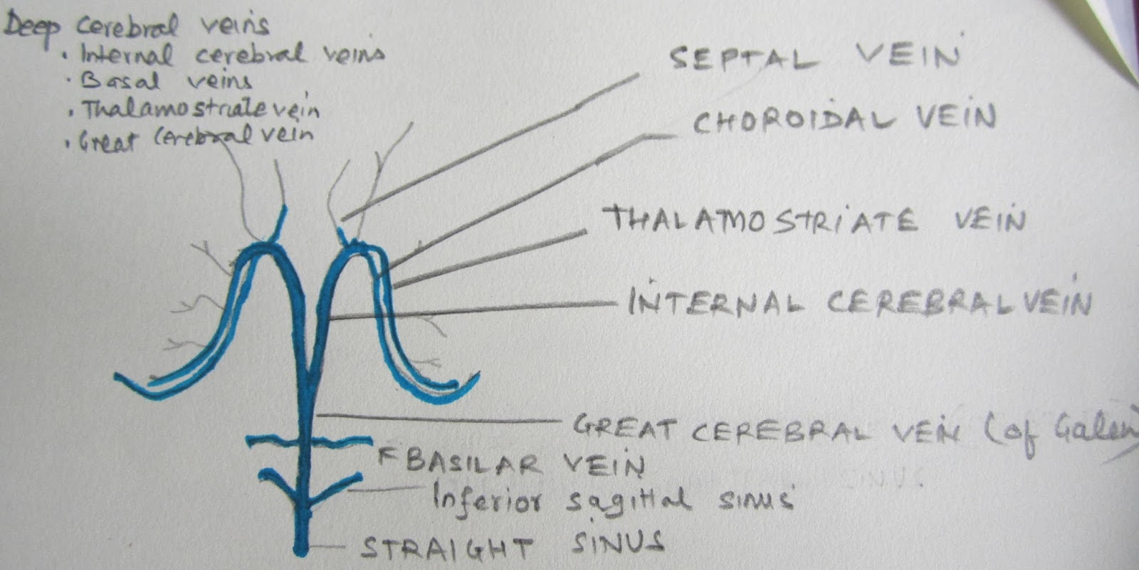
Venous system of brain

All the veins of the brain drain into Internal Jugular Vein which drains into right atrium of the heart. But the venous drainage of brain is different from other structures of the body. The veins of the brain can be described as superficial and deep venous systems of the brain.

Superficial venous system of the brain


Deep Venous System of the Brain


Cranial nerves

There are 12 pairs of cranial nerves

Cranial nerve I Olfactory nerve
Cranial nerve II Optic nerve
Cranial nerve III Oculomotor nerve
CN IV Trochlear nerve
CN V  Trigeminal nerve
CN VI Abducens nerve
CN VII Facial nerve
CNVIII Vestibulocochlear nerve
CNI X  Glossopharyngeal nrve
CN X Vagus nerve
CN XI  Spinal Acessory nerve
CN XII Hypoglossal nerve


Cranial nerves 128 are sensory nerves, 5, 7 , 9 and 10 are mixed nerves and rest are motor cranial nerves.
( So, to know the type of cranial nerves ,  remember just two numbers 128, and 5790).


Contrary to the popular belief about difficulty in neuroscience and brain anatomy, it is very easy to understand the neuroanatomy. If someone aims to master the neuroanatomy, the way is very simple. Begin with understanding the basic anatomy of skull , brain , vertebral column and spinal cord in a step wise manner. I have drawn the pictures of the skull , brain and spinal cord to highlight the value of these basic facts.

This manner of learning is good for the medical students and neurosurgeons as well. The ability to draw the image of a skull helps in understanding the parts of skull . This helps in understanding neuroradiology and in the surgical planning also.


 So, the first step is to see the skull. It is easy to identify frontal bone, parietal bone , temporal bone, occipital bone, orbit, and maxilla. locate nasal aperture in the frontal view. and pterion and zygomatic process in the lateral view of skull.


The first picture provides an idea about the three fossae of the inside of the cranium or skull. The anterior cranial fossa, middle cranial fossa and posterior cranial fossa. It is extremely important to know these three terms, as these three terms are frequently used by the experts in neuroanatomy and neurosurgery. This picture is a diagrammatic representation of the view of the skull base as seen from above after removing the skull cap.  One should learn to draw this simple yet a very important diagram.  Locate the greater wing of the sphenoid bone, anterior clinoid procees , petrous bone, posterior clinoid process and foramen magnum in this diagram. Rest of the three diagrams are are enlarged view of the base of the three intracranial fossae.

Just to make you aware about the inside of the cranium another simple diagram is drawn below . Identify and note the location of  tent which divides inside the cranium into supratentorial and infratentorial compartments.


BRODMANN'S AREAS
As you know that almost all areas of the body are represented in the cerebral hemisphere which are known as Brodmann,s areas. These areas are given different numbers. Like Motor area, Sensory area, Speech area, visual area, hearing area. 
Important functional areas of brain are : 
Primary Motor area or Primary Motor Cortex or Motor Strip or Precentral Gyrus ( area 4)          
Primary Somatosensory cortex ( areas 3,1,2 are situated in the post central gyrus, i.e. part of the Parietal lobe just posterior to the central sulcus)
Primary auditory area or Transverse gyrus of Heschl ( areas 41 & 42)                                       
    Broca's Speech Area ( area 44 )is situated in the dominant inferior frontal gyrus which is usually left inferior frontal gyrus in right handed person.
Wernicke's area ( Receptive speech area)
Frontal eye field ( area 8)                      
Primary Visual Cortex ( area 17 )

Spinal cord is the caudal continuation of the medulla oblongata. When the distal part of brain stem is continued outside the foramen magnum it becomes the spinal cord. It is contained inside the vertebral canal formed inside the vertebral column. It is bounded anteriorly by the vertebral bodies and intervertebral discs and posteriorly by the lamina and spinous processes. The spinal nerves emerge from the intervertebral foramina.

This is the schematic transverse section of the spinal cord. Spinal cord is also bounded by three meningeal layers as that of brain. Subarachnoid space contains CSF. Ligamentum denticulatum is the extension of the Piamater in spinal cord.


Anterior Cervical Discoidectomy ( ACD ) - A very important Neurosurgical Procedure.

Cervical spondylosis is a chronic degenerative process. Degenerative disease of the cervical spine is a common cause of locomotor disability in patients over the age of 50 years. Cervical spondylosis results from progressive biomechanical stress and strain and can be compounded by repetitive trauma. It involves disc degeneration and is accompanied by facet joint osteoarthritis and ossification of posterior longitudinal ligament. There is gradual decrease in viscoelasticity of the nucleus pulposus and reduction in disc volume and height.
There are various operative procedures for treating the degenerative disease of cervical spine.Various types of neurosurgical procedures can be grouped widely into two categories : Anterior and Posterior approaches.
The anterior approaches for cervical spine surgery are
1. Anterior cervical discoidectomy ( or discectomy)
2. Anterior cervical discoidectomy with grafting [autograft/ allograft ( disc prosthesis)]
3. Anterior cervical discoidectomy with grafting and plating
4. Anterior cervical corpectomy with adjacent discoidectomy with cage graft and fixation with plating
          The relevance of any of these procedures is debatable. However, there is no question about the significance of learning the surgical technique of anterior cervical discoidectomy. All neurosurgeons should know the anterior approach of surgery of degenerative disease of spine. It is easy and direct.
This procedure is performed under general anesthesia with patient in supine position and cervical spine in the neutral or very minimally extended position. Hyperextension of the neck is avoided because during surgery greater force is required to retract the tracheal and esophagus. A vertical incision along the medial border of the sternocleidomastoid on the right side is usually preferred by many neurosurgeons. Theoretically there is risk of damage to recurrent laryngeal nerve on right side but actually there is minimal risk because this nerve runs in tracheoesophageal groove. Some prefer midline horizontal incision.
After skin incision, platysma muscle is cut , then investing layer of fascia is cut and sternocleidomastoid muscle and carotid sheath is retracted laterally and trachea and esophagus is retracted medially with the Cloward retractor. Then, vertebral body is felt and prevertebral fascia is cut and retractor is applied between longus colli muscle on the anterior aspect of vertebral body. level of the disc is confirmed and discoidectomy is performed.
Closure of the surgical wound is done only in two layers- one layer of Platysma and subcutaneous tissue and then skin is closed.
So, for every neurosurgeon who wishes to specialize in spinal neurosurgery it is must to learn anterior cervical discoidectomy.
For a single level degenerative cervical disc PIVD , anterior cervical discoidectomy may be sufficient. There is lot of evidence in support of various additional procedures like Anterior cervical discoidectomy(ACD) with grafting, ACD with grafting and plating, ACD with artificial disc ( disc prosthesis), Corpectomy with adjacent discoidecomy with grafting with Plate and Screw fixation, or cage with graft with plate and screw fixation. In traumatic cervical disc prolapse fixation is advisable.
Anterior cervical discectomy with fusion ( ADCF) is regarded as the gold standard for treatment of cervical disc disease.
The anterior approaches came into practice in 1950s. Bailey & Badgley performed first anterior stabilization of the cervical spine in 1952. In 1955 Robinson and Smith described their method to stabilize cervical spine disc disease using iliac bone graft.
Cloward published his technique of anterior cervical discectomy.
The Posterior  approaches for cervical spine surgery are
Laminectomy
Laminoplasty

    These approaches are more suitable for multisegmental cervical spondylosis.


Saturday, 28 September 2013

Neuro-radiology for medical graduates

Various imaging modalities used for investigating neurological disorders are:

X-Rays ( Plain radiography)
Ultrasonography
CT scan of the brain & Spine, CT scan with contrast, CT Angiography
MRI Brain and Spine, MR Angiography, MRI with contrast, MR Spectroscopy, MR Tractography, Functional MRI of the brain, MR cisternography
Angiography, Digital substraction angiography ( DSA)
Myelography, CT myelogram
PET CT Scan
PET MRI
TCD ( Transcranial Doppler)
SPECT

Plain Radiography

X-Ray skull is useful for the diagnosis of skull bone osteomyelitis, Craniovertebral junction abnormalitise, Tumors of the cranial bones like osteomas, osteosarcoma,  metastasis to skull may be seen on skull X - ray films. Skull fractures in head injury & Growing skull fractures in children are diagnosed on skull radiography.

CT scan: CT scan is commonly used abbreviation of Computed Axial Tomography ( CAT ) scanning. This investigation machine was developed in 1970s and it was a most important development in the field of Neuroradiology after the development of X rays ( 1890s) and angiography ( 1920s and 1930s). It is a non invasive procedure and uses X-rays for the imaging. It utilizes X-Ray beam which passes through the tissue and produces a picture like x ray but in varying shades of grey.  The density of tissue changes the picture. CT scan produces axial or cross sectional ( slices) images of the body.
Computer  measures the density of the tissue through which x ray beam passes. CT scan machine uses multiple pencil beams of x ray which rotate in the gantry and pass through the body and on opposite side dosimeter measures the amount of radiation reaching it. Each cubic part of tissue is known as voxel ( in New machines about 512 voxels) . Each voxel produces a pixel. Computer measures the attenuation of the beam and assigns a Hounsfield Unit  ( HU ).
Sir Godfrey Hounsfield from England and Allan McLeod Cormack from USA shared Nobel prize in 1979 for invention of CT scan . All shades of Gray for image May be assigned a number ' HU'. Any HU value below minus 15 will appear pure black on CT film and any HU value above 155 HU will appear pure white.  Common  HU values are water zero ( 0 )
CSF in brain 10 to 16, Air minus 1000, Fat minus 60 to minus 120. Fat containing medullary bone will appear less white as compared to compact cortical bone ( HU +1000).


CT scan of the brain is the investigation of choice
For brain trauma patients, because
- it is less time consuming,
-the presence of the metal ( bullet in gun shot injury, metal in stab injury) is not a contraindication,
- an trauma patient where the history of pace maker of heart or metallic implant is not known , CT scan is possible,
-detects bony injuries, like a fracture, depressed fracture and hematoma associated with fracture
-better delineation of hematoma .
CT scan brain is also an investigation of choice
For detecting subarachnoid hemorrhage ( spontaneous subarachnoid hemorrhage) due to rupture of an intracranial aneurysm

CT angiography ( CTA ) is an investigation to detect the aneurysm of the brain. It has become an important tool for detecting the site of aneurysm bleed, location , and other characteristics of the aneurysm of the brain . It is more sensitive than MR Angiography and its sensitivity is comparable to the Digital Substarction Angiography.

CT scan of the spine: Although MRI of the spine is undoubtedly the investigation of choice for spine, CT scan of the spine is still an important investigation. CT scan of the spine is required when MRI of the spine is not possible, for example, if a patient is with metal prosthesis ( spinal instrumentation with ferromagnetic material like steel), or a metallic bullet is impinged in the spinal cord following a gun shot injury. CT spine also helps in conditions like canal stenosis, bony fractures, ossified posterior longitudinal ligaments, etc.

High resolution CT scan, 3D reconstruction, CT myelogram , Perfusion Coputed Tomography , Intraoperative CT scan are other applications of CT scan.

MRI is the most important development in the field of neuroradiology after the development of X- rays, Angiography, and CT scan.
MRI is a non invasive radiological investigation. It does not expose the patient to the risk of radiation. It uses magnetic field . It provides multiplanar images, i.e, images in sagittal, coronal and axial planes.
Functional MRI is another non invasive investigation which helps in imaging of the eloquent area of the brain.
MR Spectroscopy provides the clue about the nature of the lesion and helps in identifying infective and neoplastic lesions of the brain.
Intraoperative MRI is an advanced technique for intraoperative imaging of the lesions inside the operation theater.
How to interpret MRI brain images?
MRI images are usually black & white. There are T1 weighted, T2 weighted, FLAIR , Diffusion weighted images and if contrast is given then T1 contrast images.
To identify T1 weighted image, see the ventricles. lateral Ventricles are in the center and contain CSF. 
On CT usually only Axial images are seen but on MRI Axial, Coronal and sagittal images are seen.
On T1 weighted image, CSF will appear Black (Hypointense).
On T2 weighted image , CSF will appear White ( Hyperintense).
On FLAIR ( Flow Attenuation Inversion Recovery) the intraventricular CSF will appear Black but brain edema will appear White.
Contrast images are usually T1 contrast Images. So, CSF will appear Black and some lesions like Meningioma will become white ( Hyperintense) after contrast enhancement.


PET and SPECT are nuclear neuroimaging and help in physiological assessment. PET ( Positron Emission Tomography) is further advanced to utilize CT or MRI imaging techniques and known as PET- CT or PET-MR. PET is used for detecting metastasis and recurrence of the tumor. PET scan commonly  utilizes Flurodeoxy glucose ( FDG) which is a radioactive tracer.

Digital substraction angiography ( DSA) is invasive investigation  which involves introducing a catheter and injecting intravenous contrast into the femoral artery. It is the gold standard investigation for defining an intracranial aneurysm, Arteriovenous malformation ( AVM) , vasospasm after Subarachnoid hemorrhage ( SAH) and other diseases of intracranial vasculature.

TCD ( Transcranial Doppler ): Noninvasive investigation to detect the vasospasm in a case of SAH.
          Although Ultrasound is not a good investigation to detect intracranial pathologies as ultrasound waves do not cross bones, there are certain places where bone is very thin like temporal squama or areas in cranium which have windows like orbit. So, the flow of blood through the intracranial arteries may be detected through these windows. In vasospasm the vessels are narrowed and flow velocity increases. This is the basis of TCD, which is a noninvasive procedure and can be performed on bedside.

Ultrasonography can be used to detect hydrocephalus and meningomyelocele in prenatal period . USG can also detect hydrocephalus in infant as anterior fontanel is not closed.

Intraoperative USG is used for real time imaging , localization, extent of resection of the tumor after craniotomy at the time of neurosurgery.

Neuronavigation is used to localize the lesion, route of the surgery, safer trajectory, etc.

Neurointervention is a very promising development in the field of Neuro-radiology. It is not only useful for the diagnosis but it also offers to treat many ailments of the brain and spine. The ost important and exciting applications of neurointerventions are: Coiling of the intracranial aneurysms, Preoperative embolization of the vascular tumors like meningioma, Embolization of the intracranial and spinal AVMs, Stenting of the vessel.


Wednesday, 25 September 2013

PET ( Positron Emission Tomography) scan

A non invasive nuclear medical imaging technique which detects pair of gamma rays emitted by a positron-emitting radionuclide (tracer), which is introduced into the body ( in blood circulation ) on a biologically active molecule.The concentrations of the tracer imaged will indicate tissue metabolic activity and may indicate cancer metastasis.
The molecule most commonly used is FDG ( fluorodeoxyglucose) , an analogue of glucose. which is concentrated in the tissues of interest in an hour and at that time person is placed in the imaging scanner. The radioisotope undergoes positron emission decay ( also known as beta decay), it emits a positron, an antiparticle of electron with opposite charge. It travels for a short distance and interacts with an electron which leads to annihilation and production of a pair of gamma ( photons ) which is detected by scintillator in the scanning device.
PET scans are read alongside CT or MRI scans giving both anatomic and metabolic information. Modern PET scanners are integrated with CT scanner so called PET-CT and because two scans are performed in immediate sequence during same session , with the patient not changing position, the sets of images are precisely registered.
PET scanner is costly because of the high costs of cyclotrons needed to produce the short-lived radionuclides for the PET scanning.
The PET scan is used for both research and clinical purpose. The clinical use in neurosurgical practice is to detect metastasis and to detect recurrence of the tumor after radiotherapy and chemotherapy. For example, a patient was operated for high grade glioma about one year ago and had already received chemotherapy and radiotherapy comes to the neurosurgeon for the complaints like headache or vomiting or seizure which creates the doubt of tumor recurrence. Then the physician should advise MRI brain with contrast with MR spectroscopy and compare the pre and post operative follow up scans. Sometimes, due to redionecrosis  the distinction better tumor recurrence and radionecrosis is not very clear and in this situation a PET scan can be advised.


Chronic Subdural Hematoma

Chronic subdural hematoma fluid may be xanthochromic (yellowish) or dark brown colour. It is called chronic because duration is more than 3 ...