Lower back pain is a very common complaint in all age groups. We all have experienced back pain at some time in our life time. Sometimes, it persists and affects our routine and becomes a matter of concern. But, in fact, most of the times it is just stiffness or muscle strain due to hard work, prolong standing, sternuous excercise, play, or after lifting some heavy object. Such back pain is not due to any underlying disease. Mild or moderate level of pain gets relieved by taking analgesics (like an adult may take Diclofenac 50 mg after meal, SOS) and after taking rest. The other causes of backache are muscle sprain, muscle pull, strain, wrong posture, etc.
Pain is a subjective sensation and the complaint of backache may vary from person to person. For example, for an athlete the lower backache after a prolong run or play may not be of a great concern but similar intensity of pain may be very debilitating for an old age person.
So, mild back pain which can be explained on the basis of obvious cause like prolong walk and play should not be investigated. Bed rest is the best medical advice for relieving backache.
However, severe persistent backache without any obvious precipitating factor may warrant a consultation with the medical specialist. Orthopedicians, Physiotherapists, Sport medicine physicians, Neurologists and Neurosurgeons commonly encounter patients with complaints of lower backache.
One of the common causes of lower backache is lumbar spondylosis, which is a progressive degenerative disease of the spine. With aging the water content in the intervertebral disc, ligament and bone is gradually decreased and it restricts the movement of the spine. The vertebral column or spine consists of cervical, thoracic, lumbar and sacral part.
A model image of the entire spine
diagram of spine
Cervical part is located on the back of the neck and is the most mobile part. Even without our notice we move our neck and cervical spine for about 80,000 times in a day. So, the cervical spine is very prone for the degenerative changes in older age group. Because thoracic spine ( chest ) is relatively less mobile due to rib cage it is less prone for spondylosis. Lumbar is the lower part and it bears the weight of the body. Due to excessive weight bearing this part of spine is prone for slip disc or listhesis.
What is Lumbar Spondylosis or Lumbar Degenerative disease?
Lumbar Spondylosis is medical term to describe a degenerative disease of the
lower part of vertebral column. Our vertebral column or back bone or spine consists of four areas, namely: cervical, thoracic, lumbar and sacral region. Lower part of the spine, i.e, region below the rib cage , consists of lumar and sacral region.
There are 5 lumbar vertebrae with intervertebral discs between two adjacent vertebral bodies.So, progressive wear and tear of this region may cause different types of diseaes. Lumbar spondylosis may lead to lumbar canal
stenosis, prolapse of intervertebral disc
( PIVD) and spondylolisthesis. Lumbar spondylosis is a common cause of lower backache.
Image showing parts of the vertebral bodies with disc.
Spinal cord is contained inside the spinal column which is formed anteriorly by the vertebral bodies and discs and bounded posteriorly by the lamina and spinous processes. In this image spinal nerve roots are seen emerging from the spinal cord ( source: http://www.medicinenet.com)
Our vertebral column consists of vertebrae and the intervertebral disc which are strengthened by many ligaments. The vertebral column contains vertebral canal within this bony canal a 45 centimetre long spinal cord is contained. The spinal nerves come out through the intervertebral foramina which are bordered by disc, pedicle, vertebral body and facet joints. So, any abnormality of disc, facet joint or vertebral joint leads to narrowing of the intervertebral neural foramina which compress the spinal nerves causing pain and later neurological deficit in the form of loss of sensation and muscle weakness.
What causes Lumbar
Spondylosis?
It is due to increased
stress over the lumbar vertebrae which causes protrusion of the intervertebral
disc, calcification of the ligaments and
osteoplytes formation . Osteophytes are the abnormal bony projections. Stress
over the vertebrae is mostly due to increased body weight, sedentary lifestyle, wrong posture or due
to old age.
Due to many risk factors, the disc may age prematurely and dries up ( disc dessication ), leading to narrowing of the disc space. This in turn decreases the flexibility of the spine and osteophyte formation in the vertebral bodies.
Nerve compression causes nerve edema, alteration in nutritional transport along the nerve, and local inflammation, therefore bed rest and anti-inflammatory medications may relieve the symptoms of the patient.
Image showing a normal part of spine and part of osteoarthritic spine
What are the common symptoms of Lumbar Spondylosis?
Lumbar canal stenosis is commonly a disease of the old age and commonly occurs due to hypertrophy of the ligamentum flavum. Patient complains of pain in lower back ( Claudication ) after walking for a long distance ( Claudication distance). Gradually , over a time , this distance decreases and person starts complaining of lower backache even after walking for 100 meters. Pain gets relieved on taking rest or in sitting position. There is no pain on bending. I this way pain due to Lumbar canal stenosis differs from the pain caused by PIVD. Pain due to disc prolapse usually increases on bending.
This problem is very commonly seen in old age persons. Initially they are able to walk for a long distance with very mild pain at the end of the walk. But, gradually with advancing age the intensity of pain increases and they start feeling pain even after walking for 200 meters or so. The moment they take rest and sit idle for a moment pain subsides. So, sitting is not painful. Some people find no difficulty in cycling but prolong walking induces lower backache.
Prolapse of intervertebral disc ( PIVD)
Prolapse or buldge or protrusion of the intervertebral disc causes compression of the spinal nerve which causes lower backache. PIVD causes pain in the legs and sometimes bladder & bowel
symptoms. Pain in the
lower back is usually caused by muscle strain. It may also include sciatica
(pain that radiates from the back to the buttock and down into the leg). Onset
of pain may be immediate or occur some hours after an activity.
Pain and stiffness may be ongoing, or only occur when you
are in certain positions. The pain may get worse by coughing, sneezing,
bending, or twisting. Even sitting may induce pain. Such type of lower backache only lying on bed in certain posture may relief pain.
Image showing prolapse of the intervertebral disc posteriorly causing compression of the spinal nerve
Spondylolisthesis is another type of degeneration . The one vertebral body is slipped over the another vertebral body. It also leads to pain in lower back.
Spondylolisthesis is very common in lumbosacral region of the spine. L4 vertebra is displaced over the L5 vertebra ( L4/L5 spondylolisthesis) or L5 is displaced over the S1 verterbra ( L5/S1 spondylolisthesis). In old aged females osteoporosis is very common and the ligaments are also weakened , so the degenerative spondylolisthesis is very common in lumbosacral region.
How to recognize Lumbar Spondylosis?
One should not ignore the severe lower backache and particularly if
pain is radiating to lower limb or associated with numbness or weakness of the
lower limb. Investigations like X-ray of
the lumbosacral spine and MRI scan of
the lower back can diagnose this problem.
Sometimes, X-ray of the spine in standing posture with bending forward
& backword is required to diagnose spondylolisthesis.
MRI of the lumbosacral spine is the preferred investigation for diagnosis of cause of lower backache. It will show the alignment of the lumbar vertebrae and intervertebral disc, dural sac, lumbar canal diameter and nerve roots. So, even a minimal disc bulge is visible on MRI. MRI may exclude other causes of the lower backache line nerve sheath tumors, any other disease of this region like Potts spine ( tuberculosis), Multiple Myeloma, etc.
So, Plain X-ray and MRI of the spine are indispensable for the diagnosis of any disease of the spine. Sometimes, CT scan of the spine may be required.
What are the treatment options?
Almost everyone experiences pain after exercise or after a prolong walk or on exertion. So, all cases of mild lower backache need not to be investigated.
Treatment
will depend on the cause of the pain. Usually a strict bed rest,
lumbosacral belt and analgesics ( mild pain drugs such as aspirin, ibuprofen,
or acetaminophen) for a short period of time can relieve the symptoms.
Many treatment options are available. Physiotherapy
measures like short wave diathermy, ultrasound therapy , traction and exercise
therapy may help in chronic cases of lumbar spondylosis.
Sometimes
stronger pain relieving drugs, muscle relaxants, and drugs to reduce
inflammation may be needed.
Urgent neurosurgical intervention is required if there is high degree
of degeneration of veretebrae, or if a disk is protruded,or associated canal
stenosis or weakness or decreased sensation in the lower limbs or difficulty in
urination. Neurosurgical Microscopic discoictomy, endoscopic discoidectomy are common surgical interventions.
In
some cases, like severe spondylolisthesis, spinal surgical fixation with
implants is required.
Can
lumbar spondylosis be
prevented?
Primary prevention of lumbar spondylosis is possible by regular
exercise, physiotherapy, correct posture and weight reduction. Once
diagnosed with lumbar spondylosis a patient
should avoid lifting heavy weight & bending forward.
Posture correction, use of correct lifting techniques, avoidance of
sudden twisting movement of back, use of cushion at lower back to maintain
lumbar lordotic curve is mandatory to prevent progressive degeneration of
lumbar spine.
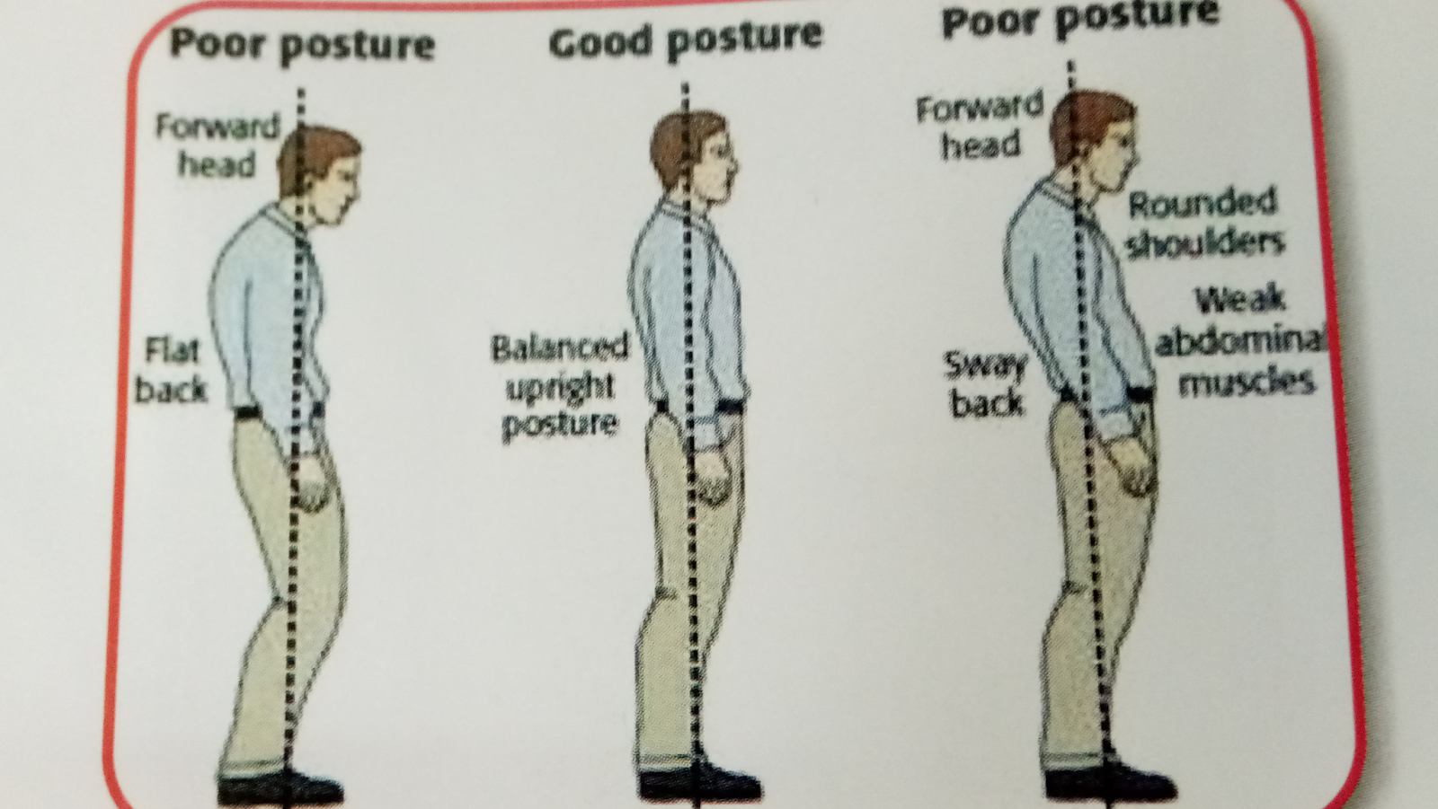
Image suggests correct standing posture standing posture without a hunch or lordosis
Surgery prevents further deterioration is severe cases. Discoidectomy and Spinal fixation may treat severe cases of spondylolisthesis and prevent further neurological deficit. Severe cases of disc extrusion should be operated to avoid foot drop and cauda equine syndrome.
Lifting of the heavy object in stooping posture is common cause of acute disc prolapse. In first image a person is lifting object in stooping posture which makes him prone to the acute neurological deficit due to PIVD. In second part the person is trying to lift the object not in stooping posture and it may be relatively safe.
Working over computer table for a long period in incorrect posture leads to early onset of spondylosis
What are the Rehabilitation options for
patients with lumbar spondylosis?
Neurological rehabilitation is a therapeutic
program designed to improve function. Various rehabilitative measure like Isometric
back exercises , core stability exercises and mobilization of lower limb joints
and stretching of tight muscles are done to maintain normal anatomical
integrity of the spine. Different interventions
like Ultrasonic therapy, Transcutaneous Electrical Nerve Stimulation ( TENS) ,
Short Wave Diathermy ,Traction and Interferential Therapy are available for
rehabilitation of the lumbar spondylosis patients.
In case of severe neurological deficit
rehabilitative therapy is available to promote compensatory strategies to
attain maximum possible functional independence. These measures are mainly
motor retraining, Neurodevelopmental
therapy ( NDT) , ADL training , Assistive and adaptive techniques and ergonomic
rehabilitation.
Other causes of lower backache like Sponylolisthesis
SPINAL TRAUMA
OSTEOPOROSIS
NERVE SHEATH TUMORS , infections of the vertebrae like spinal tuberculosis,
OSTEOARTHRITIS OF HIP JOINT
Should also be investigated.
X-ray of the lumbosacral spine ( anteroposterior and lateral view May provide an initial clue to the diagnosis.
For diagnosis of L1 vertebra collapse fracture, x- ray film of dorsolumbar spine is advised. This is very common fracture in people who fall from height.
MRI is the investigation of choice. It shows intervertebral disc, ligaments, vertebral bodies, integrity of the spinal cord.
Labelled diagram of MRI side ( sagittal view) showing different parts of lumbosacral region of spine
Sources
Naturopathy & yogic management of Lumbar spondylosis . Booklet of Central council for research in yoga and naturopathy ( An autonomous organisation under Ministry of AYUSH , Govt of India)
Online resources